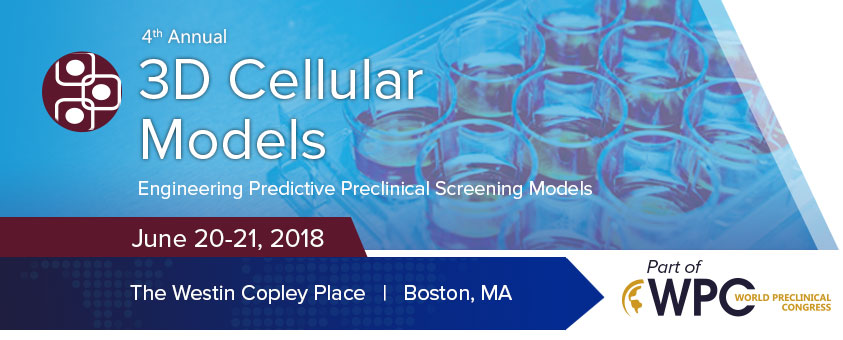
Inadequate representation of the human tissue environment during a preclinical screen can result in inaccurate predictions of a drug candidate’s effects. Thus, pharmaceutical investigators are searching for preclinical models that closely resemble
original tissue for predicting clinical outcome. Three-dimensional cell culture recapitulates normal and pathological tissue architectures that provide physiologically relevant models to study normal development and disease. However, challenges remain
for high-throughput screening as researchers must procure large numbers of identical 3D cell cultures, develop assays and obtain fast, automated readouts from these more complex assays. Join cell biologists, tissue engineers, assay developers, screening
managers and drug developers at Cambridge Healthtech Institute’s Fourth Annual 3D Cellular Models: Engineering Predictive Preclinical Screening Models conference as they discuss strategies that accelerate the identification
of novel therapeutic leads.
Final Agenda
Wednesday, June 20
11:00 am Registration Open (America Foyer)
11:45 Enjoy Lunch on Your Own
12:20 pm Dessert and Coffee Break in the Exhibit Hall with Poster Viewing (America Ballroom)
1:00 PLENARY KEYNOTE SESSION
Essex South
2:30 Refreshment Break in the Exhibit Hall with Poster Viewing (America Ballroom)
3:10 Chairperson’s Opening Remarks
Jonathan Garlick, DDS, PhD, Professor, Oral Diagnosis, School of Dental Medicine, School of Medicine and Engineering, Tufts University
3:15 KEYNOTE PRESENTATION: Drug Discovery in the Third Dimension and Beyond
 Marc Ferrer, PhD, Team Lead, NIH Chemical Genomics Center, NCATS, NIH
Three-dimensional (3D) cell cultures that mimic the spatial organization of cells in a live tissue are now being developed to test the activity of therapeutics in more predictive assay systems. Progress towards the development of an assay platform
of 3D tissue models ranging from spheroids to 3D bioprinted tissues will be presented from the perspective of a laboratory focused on drug screening and discovery.
3:45 The Massive Impact of Microtissues on Drug Discovery and Development
 Matt Wagoner, Associate Director, Mechanistic and Investigative Toxicology, Takeda Pharmaceuticals
We’ve all heard the promise of stem cell-based microtissues and organ-on-a-chip technologies – so how can they actually be used to help make better medicines? In this talk, we discuss the application of these technologies to help make
safer, more effective therapies, including Microbrains, MiniGuts and other stem cell-derived models that are transforming the way we make medicines.
4:15 Phenotypic Analysis of 3D Spheroids for Drug Screening Applications
Michael Hiatt, PhD, Senior Scientist, Research & Development, STEMCELL Technologies Inc.
Increasingly, the cancer research field is recognizing the need to transition to 3D screening approaches. However, with improved physiological relevance comes increased heterogeneity that can confound standard assay readouts and labor intensive or off-platform assays are often not desirable. We present data demonstrating the correlation of phenotypic metrics to traditional viability assays, and highlight methods for easily validating phenotypic metrics for use in establishing classifying treatment effects.
4:30 Novel 3D Assay for Immuno-Oncology and Evaluating ADCC
 Andrea Alms, Consultant, Molding Component Business Department, Kuraray Co. Ltd
Tumor microenvironment of 3D-cultures includes cell-cell interactions and formations of metabolic gradients (which are vital to understanding drug efficacy and resistance). To understand ADCC, we developed novel method to evaluate cellular toxicity
in spheroids; quantification of ADCC activity elicited by trastuzumab in 3D-culture vs 2D; and, fresh vs frozen cells.
4:45 What 3D Skin Models Teach Us: Lessons Learned from 20 Years of Building from the Ground Up
 Jonathan Garlick, DDS, PhD, Professor, Oral Diagnosis, School of Dental Medicine, School of Medicine
and Engineering, Tufts University
3D engineered tissue models of human skin have emerged as a platform technology for drug screening and modeling of human skin disease. These tissue models have evolved through integration of new understandings of stem cell biology, the cell microenvironment
and tissue engineering principles. This presentation provides an overview of what we have learned about engineering skin models over the past 20 years and what can be utilized going forward in other models.
5:05 Putting 3D Bioprinting to the Use of Tissue Model Fabrication
 Yu Shrike Zhang, PhD, Research Faculty & Associate Bioengineer, Division of Engineering
in Medicine, Department of Medicine, Brigham and Women’s Hospital, Harvard Medical School & Harvard-MIT Division of Health Sciences and Technology
The talk discusses our recent efforts on developing a series of bioprinting strategies including sacrificial bioprinting, microfluidic bioprinting, and multi-material bioprinting, along with various cytocompatible bioink formulations, for the
fabrication of biomimetic 3D tissue models. These platform technologies will likely provide new opportunities in constructing functional organoids with a potential of achieving precision therapy by overcoming certain limitations associated
with conventional models based on planar cell cultures and animals.
5:25 Engineered in vitro Disease Models for Personalized Medicine
 Gulden Camci-Unal, PhD, Assistant Professor, Chemical Engineering, University of Massachusetts
Lowell
Dr. Camci-Unal’s research aims to control and modulate cellular behavior for directing repair and regeneration of tissues. To achieve this goal, she uses diverse tools taken from chemistry, cell biology, materials science, and engineering.
She talks about new engineered biomaterial platforms to generate multicellular and compartmentalized tissue-mimetics for clinical applications including endothelialization of cardiovascular tissues, regeneration of bone, and invasion of tumors.
5:45 Close of Day and Dinner Short Course Registration*
*Separate registration required.
Thursday, June 21
7:30 am Registration Open (America Foyer) and Morning Coffee (Foyer)
8:00 Chairperson’s Remarks
Stephen S. Ferguson, PhD, Chemist, Division of the National Toxicology Program (NTP), Biomolecular Screening Branch (BSB) & NTP Laboratories, National Institute of Environmental Health Sciences (NIEHS)
8:05 Bioengineered Tissue Models of Human Development and Disease
 Shay Soker, PhD, Professor, Regenerative Medicine, Wake Forest Institute for Regenerative Medicine, Wake
Forest School of Medicine
We have created 3D human tissue model systems (organoids) that can be studied in vitro for several weeks. The organoids replicate native tissue structure and function and thus are superior to
traditional 2D cultures in order to study organ development, function and drug toxicity. Other applications focus on diseases such as tissue fibrosis and cancer, specifically, to study tumor growth and drug response for future use
in personalized/precision medicine.
8:35 Development and Application of Organotypic in vitro Liver Models to Screen Compounds for Drug-Induced Liver Injury and Transcriptomic Pathway Perturbations
 Stephen S. Ferguson, PhD, Chemist, Division of the National Toxicology Program (NTP), Biomolecular
Screening Branch (BSB) & NTP Laboratories, National Institute of Environmental Health Sciences (NIEHS)
In Phase III of Tox21 we are developing organotypic in vitro screening models to investigate chemical safety/toxicity. In this presentation, we describe our 3D HepaRG spheroid screening model (384-well), and an evaluation
of 24 reference compounds via high-throughput transcriptomics to quantitatively predict human liver injury and explore the power of resolution via concentration-response modeling to improve identification, resolution, and contextualization
of gene- and ‘pathway-level’ perturbations for translational application.
9:05 Microfabricated Organ-on-Chip Models of Tissue with High-Throughput Compatibility for Drug Screening
Joseph Charest, PhD, Program Manager, Commercial Programs, Draper
Draper’s PREDICT-96 system has 96 independent tissue replicates within a standard 96 well plate footprint, resulting in a high-throughput organ-on-chip technology. For each of the 96 independent replicates, the system has 2 microchannels
with individual pumps, a transepithelial electrical resistance (TEER) readout, and the ability to image with confocal and high-content screening systems. In addition, the system has been applied to several tissue and organ types
which will be discussed.
9:35 Find Your Table and Meet Your Moderator
9:40 Interactive Breakout Discussion Groups
This session features various discussion groups that are led by a moderator/s who ensures focused conversations around the key issues listed. Attendees choose to join a specific group and the small, informal setting facilitates sharing
of ideas and active networking.
The Use of 3D Cellular Models, Organ-on-a-Chip Technologies, or Microphysiological Systems in Preclinical Evaluation of Pharmaceuticals
William Daly, PhD, Managing Director & Faculty Scientist, Orthopedics and Rehabilitation, University of Wisconsin - Madison
- What are the key barriers to adoption of 3D cellular models in preclinical workflows?
- What are pharmaceutical wish lists (biomarkers, function, histology, etc.) for validation of 3D cellular models (groups to split into organ of choice)?
- Where is the ideal fit for 3D cellular models in the drug development pipeline / pharmaceutical testing?
- What are the missing models / unmet needs for preclinical evaluation and efficacy testing?
- Are single organs enough or is there a strong need for multi-organ testing?
3D Models of Human Organs: Challenges and Prospects
Samira Musah, PhD, Dean’s Postdoctoral Fellow, Wyss Institute at Harvard University and Harvard Medical School
- Inducing stem cell differentiation, cell maturation, and functionality
- Organoids, organs-on-chips, 3D-bioprinted tissues
- Applications in disease modeling and therapeutic discovery
Developing Academic/Industry Partnerships Using 3D Tissue Models as a Tool to Support Product Testing and Development by Start-Ups or Small Businesses
Jonathan Garlick, DDS, PhD, Professor, Oral Diagnosis, School of Dental Medicine, School of Medicine and Engineering, Tufts University
Avi Smith, MA, Senior Research Technician, Tufts University
- What value added do 3D tissue models provide as disease- or patient-specific 3D skin models for drug and product development and evaluation for early-stage businesses or start-ups?
- How can 3D skin models be used for more efficient drug development and product testing for early-stage businesses or start-ups?
- Potential financial and scientific benefits of leveraging 3D tissue models through academic partnerships
- How to develop mutually beneficial partnerships and funding sources (SBIR/STTR) with academic partners
- How to best leverage 3D tissues technologies with academic partners for commercial success
10:20 Coffee Break in the Exhibit Hall with Poster Viewing (America Ballroom)
11:05 Vascularized 3D Models of the Developing Blood-Brain Barrier and Cerebral Cortex for Developmental Neurotoxicity Screening and Disease Modeling
 William Daly, PhD, Managing Director & Faculty Scientist, Orthopedics and Rehabilitation,
University of Wisconsin - Madison
Here, we present a 3D vascularized stem cell derived organoid model (/microphysiological system) of the cerebral cortex in a microfluidics platform that has distinct neural, vascular and microglial components. The model captures
the initial vascularization of the developing brain (i.e., blood-brain barrier formation) from the perineural vascular plexus in an enhanced throughput microfluidics model. The model has been used to screen for developmental
neurotoxins and to model MeCP2 spectrum disorders.
11:35 Blood-Brain Barrier Spheroids as an in vitro Screening Platform for Brain-Penetrating Agents
 Sean Lawler, PhD, Assistant Professor & Managing Director, Harvey Cushing Neurooncology Laboratories,
Neurosurgery, Brigham and Women’s Hospital
The BBB represents a major obstacle to the delivery of drugs to the brain. We have recently developed a 3D in vitro model of the BBB, composed of astrocytes, pericytes, and endothelial
cells, which spontaneously form spheroids with BBB properties. We have identified novel peptide agents which can cross the BBB using this approach. This talk describes BBB spheroids and their capabilities as a predictive and
screening tool.
12:05 pm Neurospheroid Arrays for in vitro Studies of Alzheimer’s Disease
Daniel Irimia, MD, PhD, Associate Professor, Division of Surgery, Science & Bioengineering, Massachusetts General Hospital, Harvard Medical School, and Shriners Hospitals for Children – Boston
12:35 Networking Luncheon in the Exhibit Hall with Poster Viewing (America Ballroom)
1:55 Chairperson’s Remarks
William Daly, PhD, Managing Director & Faculty Scientist, Orthopedics and Rehabilitation, University of Wisconsin - Madison
2:00 Engineering Patient-Specific Organs-on-Chips from Human-Induced Pluripotent Stem Cells
 Samira Musah, PhD, Dean’s Postdoctoral Fellow, Wyss Institute at Harvard University and
Harvard Medical School
This talk describes our lab’s use of interdisciplinary approaches to control stem cell fate decisions and engineer functional microfluidic conduits of the human kidney glomerulus. Our most recent work involves the establishment
of a robust stem cell-based method to generate blood-filtering cells (podocytes), and integrating these cells with a microfluidic organ-on-a-chip system to recapitulate the structure, function, and specific drug toxicity of
the human kidney’s blood filtration unit.
2:30 Advanced Kidney and Liver Platforms for Drug Discovery
 Piyush Bajaj, PhD, Scientist II, Investigative Toxicology, Drug Safety Research and Evaluation,
Takeda Pharmaceuticals
In this talk, I discuss some advanced in vitro liver and kidney platforms (stem cell derived, spheroids/organoids, and organ-on-a-chip) and their applications towards different stages of drug discovery.
3:00 Application of in vitro Models to De-Risk Drug-Induced Liver Injury in Pharmaceutical Development
Wen Kang, PhD, Principal Scientist, Safety Assessment & Laboratory Animal Resources, Merck & Co., Inc.
Drug-induced liver injury (DILI) remains a major safety concern in pharmaceutical development. Implementation of physiologically relevant in vitro liver models enables the assessment of a compound's liver safety profiles
early in development. We summarize the characterization, evaluation and qualification of the micropatterned co-culture liver model HepatoPac®, and discuss how we have used the model, in conjunction with molecular
and metabolomics approaches, to inform on the DILI risk of drug candidates. Lastly, we lay out the challenges for future model development, and the next steps to refine such in vitro tools to optimize their potential.
3:30 CLOSING PANEL DISCUSSION: What Is the Biggest Bottleneck for Applying 3D Models in Discovery Research?
All agree on the potential of preclinical 3D cellular models to predict clinical outcome. However, challenges remain for high-throughput screening as researchers must procure large numbers of identical 3D cell cultures, develop
assays and obtain fast, automated readouts from these more complex assays. This panel discusses progress in:
- Standardization of the models
- Validation of the models
- Inter-laboratory reproducibility
Moderator: William Daly, PhD, Managing Director & Faculty Scientist, Orthopedics and Rehabilitation, University of Wisconsin - Madison
Panelists: Piyush Bajaj, PhD, Scientist II, Investigative Toxicology, Drug Safety Research and Evaluation, Takeda Pharmaceuticals
Jonathan Garlick, DDS, PhD, Professor, Oral Diagnosis, School of Dental Medicine, School of Medicine and Engineering, Tufts University
Wen Kang, PhD, Principal Scientist, Safety Assessment & Laboratory Animal Resources, Merck & Co., Inc.
Samira Musah, PhD, Dean’s Postdoctoral Fellow, Wyss Institute at Harvard University and Harvard Medical School
Matt Wagoner, Associate Director, Mechanistic and Investigative Toxicology, Takeda Pharmaceuticals
4:00 Close of Conference